
|
|
| Smooth | 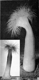 |
|
| With verrucae | 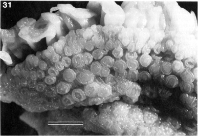 |
Figs. 30-31. Synantheopsis parulekari spec. nov. Habitus of paratype from Okha, India (RMNH Coel. 18427) and enlarged detail of its distalmost region showing prominent verrucae and marginal "pseudo-acrorhagi". Scale bars: fig. 30 = 30 mm; fig. 31 = 5 mm." Permission to display image granted by the National Natuurhistorisch Museum, Leiden, Netherlands. |
| With tenaculi | 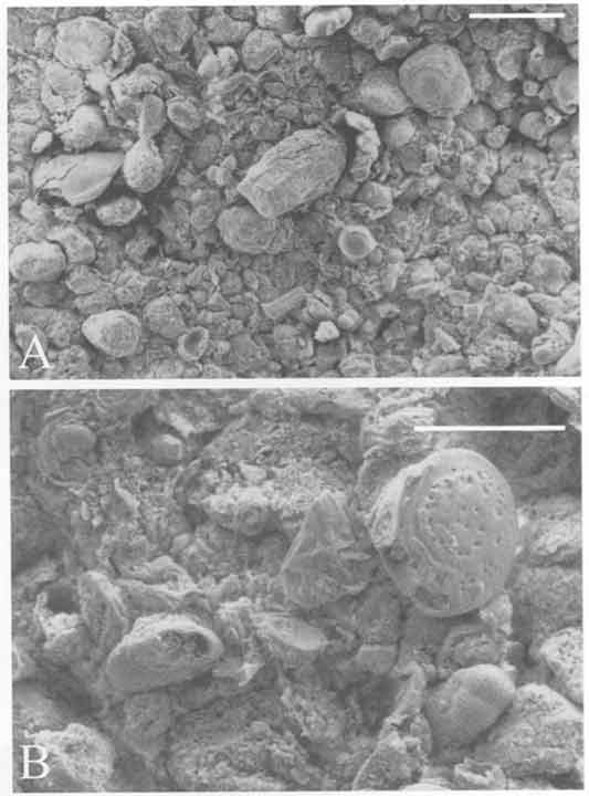 |
Fig. 6. Halcampella maxima. (A) and (B) SEM photograph of the surface of the scapus showing the tenaculi and grains of sand and foreign particles (mainly foraminifers) adhering to it. Scale bars: A, 0.5 mm; B, 0.2 mm." Permissions to display image requested from copyright holder, reply pending. |
| With vesicles | 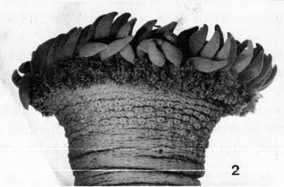 |
Saccactis coliumensis. Fig. 2. Upper half of specimen 84-01M1 showing belt of verrucae below marginal ruff of vesicles (scale bar for 1 and 2: 1 cm). Reprinted with permission of the author. |
| With marginal spherules | 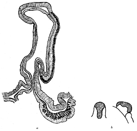 |
Fig. 17. Anthostella stephensoni n. sp. a. Section of the sphincter and of a marginal spherule. b. marginal spherule from above and from the side; dotted part violet, other parts yellow. |
| With pseudospherules | 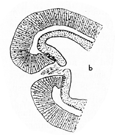 |
Fig. 2. Reprinted with permission of the Royal Swedish Academy of Sciences. |
| With tuberculate | 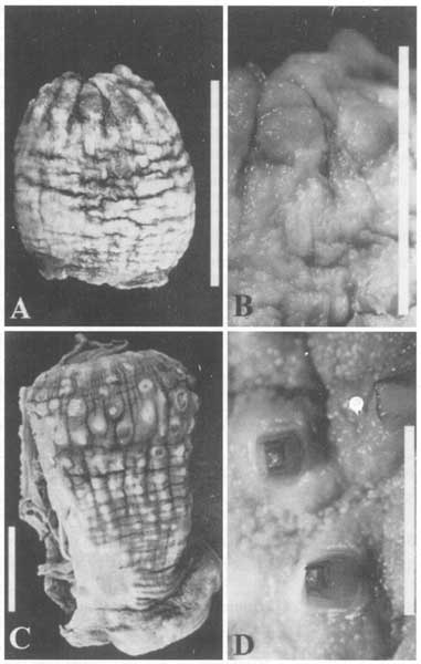 |
Fig. 6 Hormathia pectinata (Hertwig 1882) (Holotype, NHM 1889.11.25.18). (A) Lateral view of the column. (B) Detail of the rounded tubercles. Hormathia spinosa (Hertwig 1882) (Syntype, NHM 1890.7.23.1). (C) Lateral view of the column; note irregular arrangemnet of the tubercles on the scapus. (D) Detail of the pyramidal tubercles on hemispherical bases. Scale bars: A and C, 20 mm; B and D, 5 mm. Reprinted with permission of Springer-Verlag |
| With papillae | 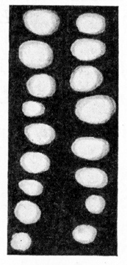 |
Fig. 1. Halcampogeton papillosus. A piece of the scapus with two rows of papillae. |
| With cinclides |  |
Figure 1. The sea anemone Acontiophorum niveum. Note the elevated cinclis (arrow). Permission to display images granted by the University of San Francisco. |
| With fosse | 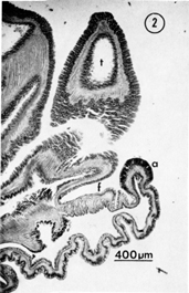 |
Fig. 2. Radial section through upper column and outer oral disc of Anthopleura handi. a = acrorhagus, f = fosse, t = tentacle. Permission to display images granted by the University of San Francisco. |
| With parapet | 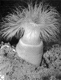 |
C-The holotype showing the collar. The invisible buried portion is about as long as the visible portion. Reprinted with permission from SIR Publishing and the Royal Society of New Zealand. |
| With spirocysts | 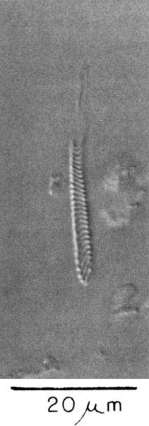 |